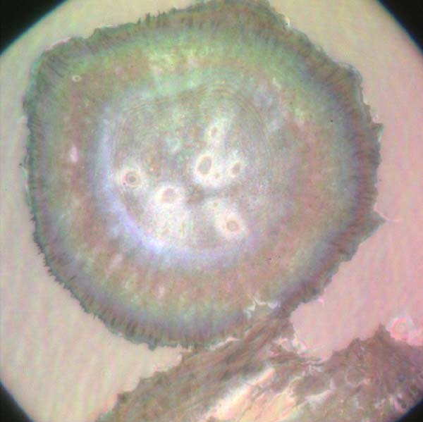
Microscopic visualization
1. Confocal Laser Scanning Microscopy using high speed spinning disk:
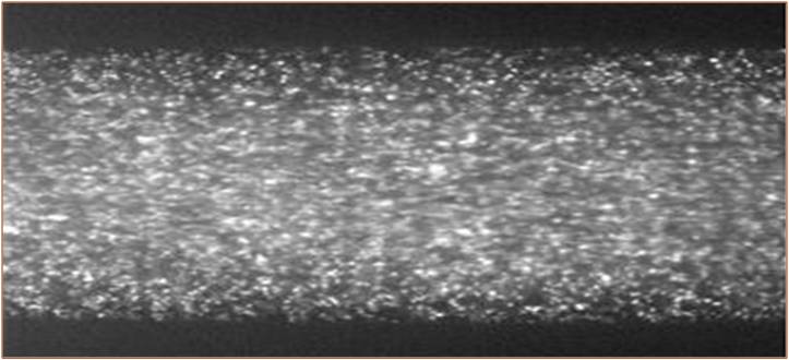
Optically sliced microscopic-particle image velocimetry (micro-PIV) is developed using confocal laser scanning microscopy (CLSM). The developed PIV system shows a unique optical slicing capability allowing true depth-wise resolved micro-PIV vector field mapping. A comparative study between CLSM micro-PIV and conventional epi-fluorescence micro-PIV is presented. Both techniques have been applied to the creeping Poiseuille flows in two different microtubes of 99-lm (Re=0.00275) and 516-lm ID diameters (Re=0.021), which are respectively
imaged by a 40?-0.75NA objective with an estimated 2.8-lm optical slice thickness, and by a 10?-0.30NA objective with a 26.7-lm slicing. Compared to conventional micro-PIV, CLSM micro-PIV consistently shows significantly improved particle image contrasts, definitions, and
measured flow vector fields agreeing more accurately with predictions based on the Poiseuille flow fields. The data improvement due to the optical slicing of CLSM micro-PIV is more pronounced with higher magnification imaging with higher NA objectives for a smaller microtube.
J.S. Park, C.
K. Choi and K. D. Kihm, Optically-Sectioned Micro PIV
Measurement Using Confocal Laser Scanning Microscopy (CLSM), ASME Journal of Heat Transfer, Vol.125,
Number 4, pp. 542, 2003
JS park, C.K. Choi,
KD Kihm, Optically
Sliced Micro-PIV Using Confocal Laser Scanning Microscopy (CLSM), Experiments in Fluids, Vol. 37, pp.
105-119, 2004
2. Optical Serial Sectioning Microscopy (OSSM):

A concept of nonintrusive thermometry is presented based on the correlation of the Brownian motion of suspended nanoparticles with the surrounding fluid temperature. Detection of fully three-dimensional Brownian motion is possible by the use of the Optical Serial Sectioning Microscopy (OSSM). This technique measures optically diffracted particle images, the so-called Point Spread Function (PSF), and determines the defocusing or line-of-sight location of the imaged particle measured from the focal plane. A dry objective lens (40X, 0.75NA) is used to detect the diffraction patterns of 500-nm polystyrene fluorescent (505/515) nanoparticles suspended in water at a volume concentration of 4x10⁻⁶, for a range of temperatures from 5 to 70?C. The measured Mean Square Displacement (MSD) data agrees fairly well with the well-known Einstein¡¯s predictions. Differentials of 5.55%, 4.26%, and 3.11% were found for 1-D, 2-D, and 3-D cases, respectively. In summary, the line-of-sight Brownian motion detection using the OSSM technique is proposed in lieu of the more cumbersome two-dimensional Brownian motion tracking on the imaging plane as a potentially more effective tool to nonintrusively map the temperature fields for nanoparticle suspension fluids.
J.S. Park, C. K. Choi and K. D. Kihm,
Temperature Measurement for Nanoparticle Suspension by detecting the Brownian
Motion Using Optical Serial Sectioning Microscopy (OSSM), Measurement
Science and Technology, Vol.16, pp. 1418-1429, 2005 [Selected as one of
the 2005 highlighted articles in MST - one of the very best contributions of
the last year, and have received the highest praise from the Board and referees
alike, whilst also being the most highly-downloaded articles throughout 2005]
3. Total Internal Reflection Fluorescence Microscopy (TIRFM):
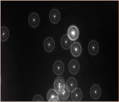
a) ( = : non-TIRF conditions), (b) and 271.6 nm evanescent wave field penetration depth ( : TIRF conditions), and (c) Changing incident angles from to . The penetration depth of is 80.7 nm.
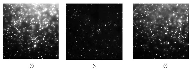
Multi-layered distributions of hindered mean square displacement (MSD) for nanoparticles are measured in the near-wall region within 500-nm from the solid surface using Total Internal Reflection Fluorescence Microscopy (TIRFM), an evanescent wave microscopic imaging technique. Examined particles are yellow-green (505/515) polystyrene fluorescent nanospheres of 100, 250, and 500 nm radii with a specific gravity of 1.055. To ensure the measurement accuracy, special care is taken to minimize photobleaching of fluorescent particles by adding neutral density (ND) filters to optimally reduce the excitation power. The experimental results for parallel MSD¡¯s to the solid surface validate the theory of hindered diffusion7 of spheres based on viscous slow-down in the near-wall region. It is also reported that the effect of adding sodium chloride up to 10 mM to the solution has little effect on the parallel diffusive motion of the tested nanoparticles. Experimental evidence shows that normal MSD¡¯s, for submicroscopic charged nanoparticles, are substantially different than that predicted by Brenner¡¯s theory8. It should be mentioned however that Brenner¡¯s work has been proven when electrostatic forces are negligible and thus there should be no implication that the theory itself is incorrect.
C.K. Choi, C.
H. Margraves, and K. D. Kihm, Examination of Near-wall hindered
Brownian diffusion of nanoparticles: Experimental validation of theories
by Brenner (1961) and Goldman et al. (1967), Physics of fluids, Vol. 19, Issue 10, 103305, 2007
C. H. Margraves, C. K. Choi,
and K. D. Kihm, Examination of the effect of
salinity on the minimum elevation of nano-particles using ratiometric
total internal reflection fluorescence microscopy (R-TIRFM), Experiments
in fluids, Vol. 41, pp. 173-183, 2006
K. D. Kihm,
A. Benerjee, C. K. Choi, and T. Takagi, Near-Wall
Hindered Brownian Diffusion of Nanoparticles Examined By Three-Dimensional
Ratiometric Total Internal Reflection Fluorescence Microscopy (3D
R-TIRFM), Experiments in Fluids,
Vol.37, Issue 6, pp. 811-824, 2004
4. Epi-fluorescence Microscopy (EFM):
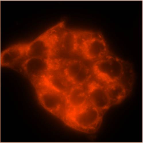
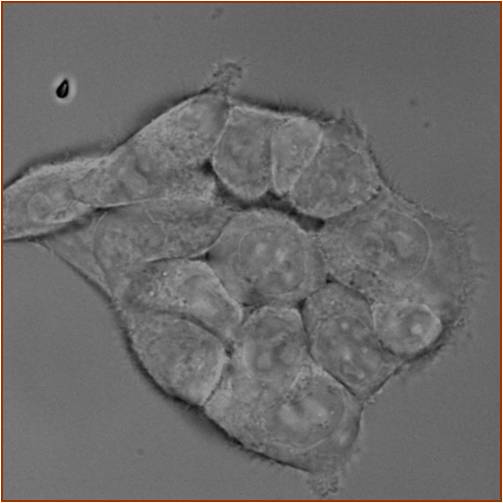
Epi-fluorescence Microscopy can be widely used for measuring fluorescence particles¡¯ motion in any fluids and tracking green fluorescence protein (GFP) in live cells and this microscopic technique is comprehensively and effectively combined with other microscopic techniques such as total internal reflection microscopy, confocal microscopy, differential interference contrast microscopy, and so on.
5. Phase Contrast Microscopy (PCM):

In phase specimens, the direct zeroth order light passes through or around the specimen undeviated. However, the light diffracted by the specimen is not reduced in amplitude as it is in a light-absorbing object, but is slowed by the specimen because of the specimen's refractive index or thickness (or both). This diffracted light, lagging behind by approximately 1/4 wavelength, arrives at the image plane out of step (also termed out of phase) with the undeviated light but, in interference, essentially undiminished in intensity. The result is that the image at the eyepiece level is so lacking in contrast as to make the details almost invisible.
6. Differential Interference Contrast Microscopy (DICM):
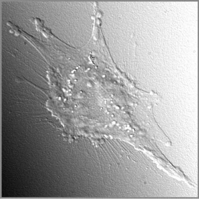
The wave pairs employed in differential interference contrast are generated by the action of a birefringent beamsplitter (Nomarski compound prism) on a plane-polarized wavefront of coherent light originating from a light soruce and focused into the front focal plane of the microscope condenser (where the beamsplitter is positioned). After wavefronts generated by the beamsplitting prism pass through a specimen phase gradient, they are recombined through differential interference by a second prism and an analyzer (another polarizer) to yield a high-contrast rendition of the gradient.
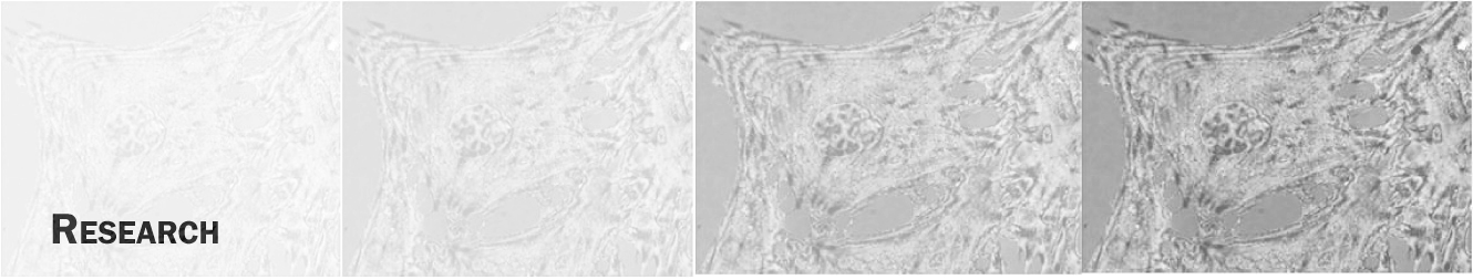
This study quantifies the dynamic attachment and spreading of porcine pulmonary artery endothelial cells (PPAECs) on optically thin, indium tin oxide (ITO) biosensors using simultaneous differential interference contrast microscopy (DICM) and electrical micro-impedance spectroscopy. A lock-in amplifier circuit monitored the impedance of PPAECs cultivated on the transparent ITO bioelectrodes as a function of frequency between 10 Hz and 100 kHz and as a function of time, while DICM images were simultaneously acquired. A digital image processing algorithm quantified the cell covered electrode area as a function of time. The results of this study show that the fraction of the cell covered electrode area is in qualitative agreement with the electrical impedance during the attachment phase following cell settling on the electrode surface. The possibility of several distinctly different states of electrode coverage and cellular attachment giving rise to similar impedance signals is discussed.
7. Interference Reflection Contrast Microscopy (IRCM):
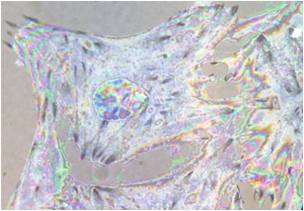
The monochromatic IRM images can be obtained by using one of RGB filters while the color IRM uses all of them, which provides higher measurement resolutions because of its multiplicity in fringe spectra. Thus, the two types of IRM complement each other and can be used simultaneously to more completely map the cell-substrate separation distribution, i.e., the former measures the cell-substrate separation with higher signal-to-noise ratios while the latter determines the slope of the separation topology. monochromatic fringes are constructed by the two reflected rays respectively from the substrate-medium and medium-cell interfaces, allowing measurement of the gap at an accuracy of ~ 100 nm, for the case of a green filter. Colored fringes are constructed with RGB filters and they are constructed in different orders depending on the slope of the separation distance topology, i.e., the spectrum orders in R-B-G-R for a positive slop and R-G-B-R for a negative slope. Simultaneously using monochromatic and color IRM measurements and a direct visualization of the cytoskeleton, this study will investigate the field distribution of cell-matrix height and focal adhesion densities.
Multi-contrast microscopy techniques were used to comprehensively and dynamically map the cellular contact area adhering to a substrate. The natural fringe patterns observed with interference reflection contrast microscopy were used to map the dynamic fingerprint of a porcine pulmonary artery endothelial cell¡¯s ventral surface and to examine the focal and/or close contacts to the substrate, when exposed to a toxic agent Cytochalasin D. In addition, differential interference contrast microscopy sequentially imaged the overall cellular morphological responses to the agent. It was observed that focal contacts, which are tightly attached to the substrate, are strongly resistant to even high doses of the cytotoxic agent and that they also form the basis of cellular recovery after replacement of the cytotoxic medium with fresh medium.
C.K.
Choi, K. Kihm, and A. English, Opto-electric
indium-tin-oxide (ITO) biosensor for simultaneous cellular imaging and
micro-impedance analyses, Optics
Letters, Vol. 32, Issue 11, pp1405-1407, 2007
C.K. Choi, A. English, K. Kihm, and C Margraves, Dynamic
Optical and Electrical Properties of Endothelial Cell Attachment on Indium
Tin Oxide Bio-electrodes, Journal of Biomedical Optics Vol. 12, Issue 6, 064028, 2007
C. K. Choi, C. H. Margraves, K. D. Kihm, and A.E. English, Dynamic Fingerprinting of
Cellular Focal Contacts under Cytotoxic Conditions Using Quantitative Analysis
of Natural Fringes, Journal
of Biomedical Optics, Vol. 13, Issue 5, 054069, 2008